Scientists get new look at how the brain processes visual information
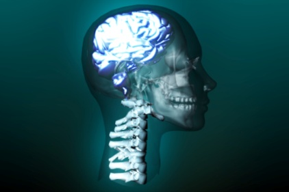 Every time you open your eyes, visual information flows into your brain, which interprets what you’re seeing. Now, for the first time, MIT neuroscientists have noninvasively mapped this flow of information in the human brain with unique accuracy, using a novel brain-scanning technique.
This technique, which combines two existing technologies, allows researchers to identify precisely both the location and timing of human brain activity. Using this new approach, the MIT researchers scanned individuals’ brains as they looked at different images and were able to pinpoint, to the millisecond, when the brain recognizes and categorizes an object, and where these processes occur.
Until now, scientists have been able to observe the location or timing of human brain activity at high resolution, but not both, because different imaging techniques are not easily combined. The most commonly used type of brain scan, functional magnetic resonance imaging (fMRI), measures changes in blood flow, revealing which parts of the brain are involved in a particular task. However, it works too slowly to keep up with the brain’s millisecond-by-millisecond dynamics.
Another imaging technique, known as magnetoencephalography (MEG), uses an array of hundreds of sensors encircling the head to measure magnetic fields produced by neuronal activity in the brain. These sensors offer a dynamic portrait of brain activity over time, down to the millisecond, but do not tell the precise location of the signals.
The MIT researchers are the first to use it to link fMRI and MEG data from human subjects.
In the study, the researchers scanned 16 human volunteers as they looked at a series of 92 images, including faces, animals, and natural and manmade objects. Each image was shown for half a second.
Each subject underwent the test multiple times — twice in an fMRI scanner and twice in an MEG scanner — giving the researchers a huge set of data on the timing and location of brain activity.
By analyzing this data, the researchers produced a timeline of the brain’s object-recognition pathway very similar to results previously obtained by recording electrical signals in the visual cortex of monkeys, a technique that is extremely accurate but too invasive to use in humans.
About 50 milliseconds after subjects saw an image, visual information entered a part of the brain called the primary visual cortex, or V1, which recognizes basic elements of a shape, such as whether it is round or elongated. The information then flowed to the inferotemporal cortex, where the brain identified the object as early as 120 milliseconds. Within 160 milliseconds, all objects had been classified into categories such as plant or animal.
The research was funded by the National Eye Institute, the National Science Foundation, and a Feodor Lynen Research Fellowship from the Humboldt Foundation.
Source: MIT
Looking for a reprint of this article?
From high-res PDFs to custom plaques, order your copy today!